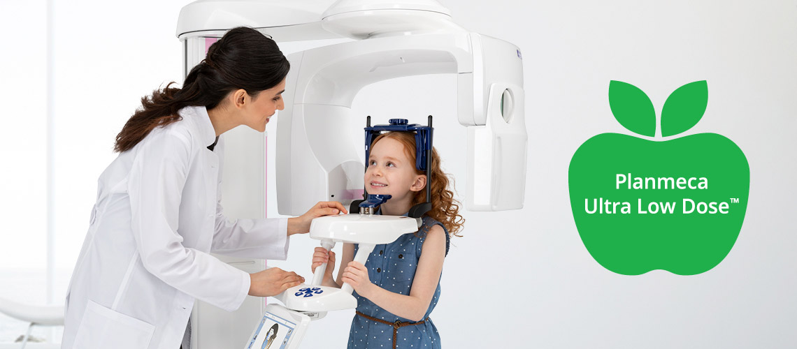
Scientifically proven low dose imaging
Planmeca’s unique Planmeca Ultra Low Dose™ 3D imaging protocol offers a scientifically proven method for low dose CBCT imaging, helping clinicians everywhere to adhere to the ALADA (As Low As Diagnostically Acceptable) principle. While using the protocol decreases the exposure values and thus the patient dose, the image quality is kept on a diagnostically acceptable level. The method provides an optimal balance between the image quality and patient dose, making it ideal for a wide range of clinical cases from implant planning to orthodontics. The use of Planmeca Ultra Low Dose protocol and its benefits have been studied and scientifically proven in various scientific research.

Planmeca Ultra Low Dose offers the best balance between image quality and dose for both small and large FOVs
EzEldeen et al. evaluated the Planmeca Ultra Low Dose protocols of a Planmeca ProMax 3D unit along with other low-dose protocols. According to the study, the Planmeca protocol had the best balance between image quality and dose for both large and small field of views.
The study concluded that applying dose optimisation protocols allows a considerable reduction in the effective dose (ED) while maintaining sufficient image quality for CBCT-based 3D planning, 3D printing of tooth replica, and post-operative follow-ups.
Source: EzEldeen, M., Stratis, A., Coucke, W., Codari, M., Politis, C., Jacobs, R. (2016). As Low Dose as Sufficient Quality: Optimization of Cone-beam Computed Tomographic Scanning Protocol for Tooth Autotransplantation Planning and Follow-up in Children. Journal of Endodontics, 43(2). https://doi.org/10.1016/j.joen.2016.10.022
A considerable reduction in the pediatric ED can be achieved while maintaining sufficient image quality for tooth autotransplantation planning and follow-up using the dose optimization protocols
Compared with standard protocols, Planmeca Ultra Low Dose provides comparable diagnostic information with a significantly smaller dose
Ihlis R.L. et al. studied the overall image quality and evaluated the visibility of anatomic structures on low-dose CBCT scans. The study concluded that images captured with high-definition and standard Planmeca Ultra Low Dose protocols (ULDHD and ULD, respectively) were diagnostically acceptable and can be recommended for the assessment of impacted maxillary canines.
Source: Ihlis, R.L., Kadesjö N., Tsilingaridis G., Benchimol D. & Shi X.Q. (2022). Image quality assessment of low-dose protocols in cone beam computed tomography of the anterior maxilla. Oral Surgery, Oral Medicine, Oral Pathology and Oral Radiology, 133(4), 483–491. https://doi.org/10.1016/j.oooo.2021.10.001
ULDHD, ULD, and LDHD protocols may be recommended for clinical studies on assessing impacted maxillary canines because these protocols provide comparable diagnostic information with a radiation dose of 23% to 39% of the standard protocol recommended by the manufacturer.
Planmeca Ultra Low Dose and Planmeca ProMax 3D preserve diagnostic image quality for evaluating fine anatomical structures
Charuakkra et al. compared the image quality and effective doses between the low dose and standard protocols of different CBCT imaging units. The overall effective doses for the low dose protocols were approximately six times lower compared to standard protocols. At the same time, while the radiation dose levels decreased, the diagnostic image quality necessary for evaluating delicate dentomaxillofacial anatomy was preserved when using the Planmeca ProMax 3D imaging unit.
Additionally, the study concluded that the subjective image quality was not significantly different between the low dose and standard protocols with a high voltage tube (120 kV). The Planmeca ProMax 3D unit with a 120 kV tube also had the best observer reproducibility.
Source: Charuakkra, A., Mahasantipiya, P., Lehtinen, A., Koivisto, J., Järnstedt, J. (2022). Comparison of subjective image analysis and effective dose between low-dose cone-beam computed tomography machines. Dentomaxillofacial Radiology. https://doi.org/10.1259/dmfr.20220176
High-tube-voltage protocols could remarkably reduce the imaging dose without degrading the image quality. Specifically, ULD and LD CBCT protocols may be adopted as routine practice for diagnosis and treatment planning.
Ultra Low Dose CBCT scans clinically sufficient for imaging the temporal bone area
Tamminen et al. evaluated the clinical quality of CBCT images taken with the Ultra Low Dose protocol for imaging the temporal bone area. The ULD images were compared to high resolution scans, with the conclusions stating that the image quality of the ULD CBCT scans was clinically sufficient.
Source: Tamminen, P., Järnstedt, J., Lehtinen, A. et al. Ultra-low-dose CBCT scan: rational map for ear surgery. Eur Arch Otorhinolaryngol (2022). https://doi.org/10.1007/s00405-022-07592-4
Using ultra-low doses of radiation, the produced IQ is clinically sufficient. We encourage ear surgeons to check the patients’ imaging history and to consider the use of imaging modalities that involve lower radiation doses especially when conducting repetitive investigations and with children.
Reducing patient doses in pre-implant assessment with Planmeca Ultra Low Dose
The in vitro study by Liljeholm et al. evaluated the overall image quality as well as the visibility of most anatomical structures and bone quality assessment in images that were captured using Planmeca Ultra Low Dose protocol. The high- and mid-definition protocols (UL-HD and UL-MD, respectively) were found diagnostically acceptable for pre-implant radiographic assessment.
Source: Liljeholm, R., Kadesjö, N., Benchimol, D., Hellén-Halme, K. & Xie-Qi, S. (2017). Cone-beam computed tomography with ultra-low dose protocols for pre-implant radiographic assessment: An in vitro study. European Journal of Oral Implantology, 10(3), 351–359. https://www.researchgate.net/publication/320585393
Low-dose protocols may be applied for pre-implant radiographic assessment. For the CBCT unit ProMax 3D Classic, the UL-HD and UL-MD protocols were preferred to the GS protocol for radiographic assessment prior to implant surgery, due to a reduction of radiation to the patient.
No statistical reduction in image quality between Planmeca Ultra Low Dose and standard protocols
The in vitro study by Ludlow and Koivisto evaluated effective doses and compared Planmeca Ultra Low Dose protocol with standard exposures. In the study, no statistical reduction in image quality was found between the Planmeca Ultra Low Dose and standard protocols.
Source: Ludlow, J. B. & Koivisto, J. (2015). Dosimetry of Orthodontic Diagnostic FOVs Using Low Dose CBCT protocol. https://www.planmeca.com/ULD-poster
An average reduction in dose of 77% was achieved using ULD protocols when compared with standard protocols. While this dose reduction was significant, no statistical reduction in image quality between ULD and standard protocols was seen.
Orthodontic cephalometric measurements benefit from Planmeca Ultra Low Dose
The study by van Bunningen et al. compared lateral cephalograms (LC) reconstructed from Planmeca Ultra Low Dose CBCT scans with traditional standard-dose lateral cephalograms to evaluate and compare the orthodontic diagnostic measurements. According to the research, there were no significant differences between the standard-dose and ultra low dose-low dose (ULD-LD) images, although the patient doses with Planmeca ULD-LD protocol were considerably lower.
Source: van Bunningen, R.H., Dijkstra, P. U., Dieters, A., van der Meer, W. J., Kuijpers-Jagtman, A. M. & Ren, Y. (2021). Precision of orthodontic cephalometric measurements on ultra low dose-low dose CBCT reconstructed cephalograms. Clinical Oral Investigations, 26, 1543–1550. https://doi.org/10.1007/s00784-021-04127-9
Based on the lower radiation dose and the small differences in variation in cephalometric measurements on reconstructed LC compared to standard dose LC, ULD-LD CBCT with reconstructed LC should be considered for orthodontic diagnostic purposes.
Planmeca ULD protocol can be applied for the detection of MB2 maxillary molars
Ahmed investigated how the Planmeca Ultra Low Dose protocol compares to a normal protocol in detecting the second mesiobuccal root canal in maxillary molars. The tests were carried out with three different voxel sizes (75 μm, 100 μm and 200 μm).
The study found that there was no statistically significant difference between the two protocols for the same voxel size and concluded that the Planmeca Ultra Low Dose protocol can be applied for the detection of MB2 maxillary molars.
Source: Ahmed, D.F. (2019). Ultra-low-dose versus normal-dose scan protocol of Planmeca ProMax 3D Mid CBCT machine in detection of second mesiobuccal root canal in maxillary molars: an ex vivo study. Egyptian Dental Journal. https://edj.journals.ekb.eg/article_71405.html
The ULD CBCT protocol can be applied for the detection of MB2 of maxillary molars. The smaller the voxel size, the higher the image resolution and image quality. The 0.075 mm voxel size of both protocols is accurate enough to be used as a gold standard in laboratory studies instead of the standard root sectioning technique.
Planmeca Ultra Low Dose evaluated as the preferred exposure protocol for CBCT images prior and after root canal treatments
In the in vitro study by Yeung et al., the subjective image quality of different CBCT exposure protocols for endodontic indications were evaluated by twelve professionals – 4 endodontists, 4 periodontists and 4 radiologists. The Planmeca Ultra Low Dose scans got the highest grades in the tests. The study concluded that a low-dose protocol did not seem to affect the perception of image quality. Furthermore, the clinical findings demonstrated that a low-dose CBCT mode could even have potential for diagnostics prior to or following endodontic treatments.
Source: Yeung, A. W. K., Harper B., Zhang, C., Neelakantan, P. & Bornstein, M. M. (2020). Do different cone beam computed tomography exposure protocols influence subjective image quality prior to and after root canal treatment? Clinical Oral Investigations, 25, 2119–2127. https://doi.org/10.1007/s00784-020-03524-w
Based on the present in vitro data, a low-dose CBCT mode seems not to negatively affect the perception of image quality.
Planmeca Ultra Low Dose protocol is available for all Planmeca CBCT imaging units as a standard feature and can be used with all volume sizes.
